

Published on Sep 12, 2023
The genetic information of plants, animals and other eukaryotic organisms resides in several (or many) individual DNA molecules, or chromosomes. For example, each human cell possesses 46 chromosomes, while each cell of an onion possesses 8 chromosomes. All cells must replicate their DNA when dividing. During DNA replication, the two strands of the DNA double helix separate, and for each original strand a new complementary strand is produced, yielding two identical DNA molecules. DNA replication yields an identical pair of DNA molecules (called sister chromatids) attached at a region called the centromere. DNA replication in eukaryotes is followed by the process called mitosis which assures that each daughter cell receives one copy of each of the replicated chromosomes. During the process of mitosis, the chromosomes pass through several stages known as prophase, metaphase, anaphase and telophase. The actual division of the cytoplasm is called cytokinesis and occurs during telophase. During each of the preceding stages, particular events occur that contribute to the orderly distribution of the replicated chromosomes prior to cytokinesis.
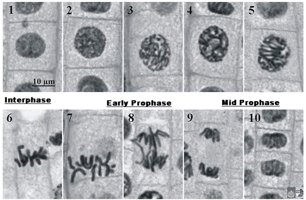
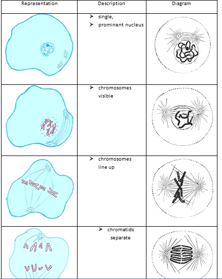
During prophase, the chromosomes supercoil and the fibers of the spindle apparatus begin to form between centrosomes located at the pole of the cells. The nuclear membrane also disintegrates at this time, freeing the chromosomes into the surrounding cytoplasm.
During prometaphase, some of the fibers attach to the centromere of each pair of sister chromatids and they begin to move toward the center of the cell.
At metaphase the chromosomes have come to rest along the center plane of the cell.
During anaphase, the centromeres split and the sister chromatids begin to migrate toward the opposite poles of the cell.
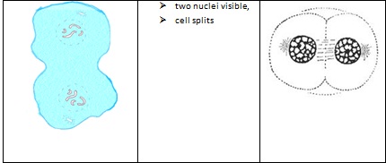
During telophase, the chromosomes at either end of the cell cluster begin to cluster together, which facilitates the formation of a new nuclear membrane. This also is when cytokinesis occurs, leading to two separate cells. One way to identify that telophase has begun is by looking for the formation of the cell plate, the new cell wall forming between the two cells.
• Better understand the process and stages of mitosis.
• Prepare your own specimens of onion root in which you can visualize all of the stages of mitosis.
• Apply an analytical technique by which the relative length of each stage of mitosis can be estimated
Small fresh onion
Small jars, glasses, or beakers
Toothpicks
Muriatic acid (10% HCl) found at hardware stores
Safety goggles
Latex gloves
Forceps or tweezers
Distilled water
Pipette or eye dropper
Paper towels
Razor blade or scalpel
2 needles or pins
0.5% Toluidine blue available online
Microscope slides and coverslips
Dissecting microscope or magnifying lens
Compound light microscope
Create your hypothesis as to which phase of mitosis you believe will be most prevalent in your sample and why. Base this hypothesis on which phase you believe will take the longest amount of time in the cell. Next, insert 3-4 toothpicks around the sides of the onion and place the onion root side down (stem side up) in a glass or jar of water. The bottom of the onion should be submerged in the water. Wait a few days for roots to grow.
Once there are long roots growing on the onion, remove the onion from the water and use the razor blade or scalpel to slice about 5 mm off the tips of the roots. Place the root tips into another small beaker or thin-walled glass jar. Pour muriatic acid into the jar with the root tips. Wear hand and eye protection while working with the acid. Allow the root tips to sit in the acid for about 20 minutes.
Use forceps to remove the root tips from the acid and rinse the acid down the drain with plenty of running water. Place the root tips on a microscope slide and rinse them several times with a pipette and distilled water. Wear hand and eye protection during these steps as well.
Carefully use the razor or scalpel to trim the root tips to 2 mm long, keeping the tips. Use needles or pins to carefully slice the root tips into 2 or more length-wise sections. Doing this work with a magnifying lens or under a dissecting microscope may be helpful.
Use a pipette to coat the dissected root tips in Toluidine blue. Let sit for 2 minutes. Place a coverslip on top and pipette distilled water onto the slide. Use a paper towel to soak up excess dye. Use more than one microscope slide for multiple roots, with only about 2 root tips per slide.
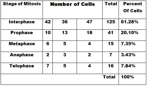
Place the slide on the microscope. Turn the microscope on and make sure that it is placed on its lowest magnification first. Focus the image until you can clearly distinguish the tip of the root. Move up slightly from the tip and focus on the area just above root tip where the cells will be replicating. Change the microscope objective to the next highest magnification. Focus on the area of cell replication. Identify the different stages of mitosis. If this is difficult at this magnification, try moving up to the highest magnification. Once you are able to easily identify the stages of mitosis within the area of cell replication, draw a sketch of each stage that you view. Compare the sketches to internet or textbook images of the stages to ensure that you are correctly identifying each stage of mitosis. Now that you are able to easily and correctly identify each stage, begin counting the cells in each stage. You must do this carefully and systematically to avoid counting cells more than once. Try moving from one area of the root tip, down and over to another area. Count many cells in multiple root tips, with a goal of counting at least 1000 cells. Take breaks between root tips if needed to rest your eyes and recheck your sketches. Record the number of cells in each stage with tick marks in a table. Having someone help you record the numbers while you count might be helpful.
Once you have counted at least 1000 cells, convert your tick marks to numbers. Graph your results with a bar graph. Compare your results to your hypothesis and draw conclusions about which stage of mitosis is most common and takes the longest amount of time.
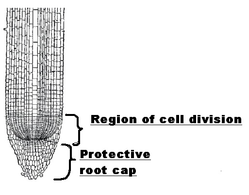
Scan the microscope under the 10x objective. Look for the region that has large nuclei relative to the size of the cell; among these cells will be found cells displaying stages of mitosis. Examples are shown in the figure to the right. Switch to the 40X objective to make closer observations. Since prophase and prometaphase are difficult to distinguish, classify all these cells as prophase. Record your observations in the table provided.
Now that we are able to identify cells in different stages of the cell cycle, now it is time to understand how long a cell will tend to be at each of these stages.
This is to count how many cells in your root tip specimen were ‘frozen in time’ in different stages of division when this slide was made.
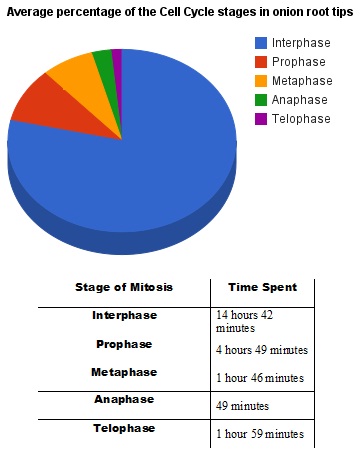
While making your observations, consider the relative number of cells actually involved in mitosis. Some of these cells are still involved in the cell cycle, which encompasses all of the processes involved in cell replication. Cell that are actively dividing but not yet in mitosis are said to be in interphase, during which time the DNA is copied and the cell is otherwise preparing for replication. Some root cells have ceased dividing and are only increasing in size, whereas others have reached their final, mature size and function, and are said to be in the GO stage.
http://www.biology.arizona.edu/cell_bio/activities/cell_cycle/cell_cycle.html
http://www.marietta.edu/~biol/introlab/Onion%20root%20mitosis.pdf
http://www.biologycorner.com/worksheet/mitosis_onion_key.html