Published on Feb 13, 2025
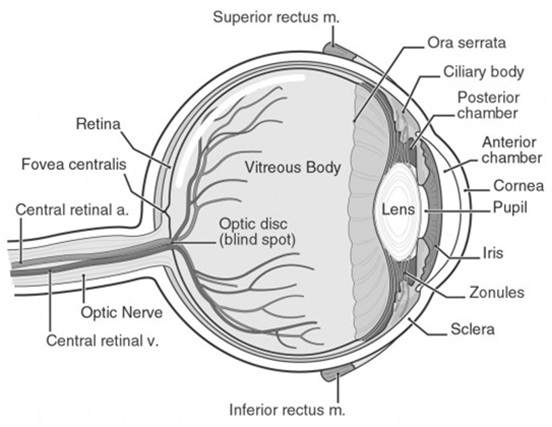
The human eye is an organ which reacts to light and pressure. As a sense organ, the mammalian eye allows vision. Human eyes help provide a three dimensional, moving image, normally colored in daylight. Rod and cone cells in the retina allow conscious light perception and vision including color differentiation and the perception of depth. The human eye can differentiate between about 10 million colors and is possibly capable of detecting a single photon. Similar to the eyes of other mammals, the human eye's non-image-forming photosensitive ganglion cells in the retina receive light signals which affect adjustment of the size of the pupil, regulation and suppression of the hormone melatonin and entrainment of the body clock.
The eye is not shaped like a perfect sphere, rather it is a fused two-piece unit, composed of the anterior segment and the posterior segment. The anterior segment is made up of the cornea, iris and lens. The cornea is transparent and more curved, and is linked to the larger posterior segment, composed of the vitreous, retina, choroid and the outer white shell called the sclera. The cornea is typically about 11.5 mm (0.3 in) in diameter, and 1/2 mm (500 um) in thickness near its center. The posterior chamber constitutes the remaining five-sixths; its diameter is typically about 24 mm. The cornea and sclera are connected by an area termed the limbus. The iris is the pigmented circular structure concentrically surrounding the center of the eye, the pupil, which appears to be black. The size of the pupil, which controls the amount of light entering the eye, is adjusted by the iris' dilator and sphincter muscles Light energy enters the eye through the cornea, through the pupil and then through the lens.
The lens shape is changed for near focus (accommodation) and is controlled by the ciliary muscle. Photons of light falling on the light-sensitive cells of the retina (photoreceptor cones and rods) are converted into electrical signals that are transmitted to the brain by the optic nerve and interpreted as sight and vision.
There are many diseases, disorders, and age-related changes that may affect the eyes and surrounding structures. As the eye ages, certain changes occur that can be attributed solely to the aging process. Most of these anatomic and physiologic processes follow a gradual decline. With aging, the quality of vision worsens due to reasons independent of diseases of the aging eye. While there are many changes of significance in the non-diseased eye, the most functionally important changes seem to be a reduction in pupil size and the loss of accommodation or focusing capability (presbyopia).
The area of the pupil governs the amount of light that can reach the retina. The extent to which the pupil dilates decreases with age, leading to a substantial decrease in light received at the retina. In comparison to younger people, it is as though older persons are constantly wearing medium-density sunglasses. Therefore, for any detailed visually guided tasks on which performance varies with illumination, older persons require extra lighting. Certain ocular diseases can come from sexually transmitted diseases such as herpes and genital warts. If contact between the eye and area of infection occurs, the STD can be transmitted to the eye.
With aging, a prominent white ring develops in the periphery of the cornea called arcussenilis. Aging causes laxity, downward shift of eyelid tissues and atrophy of the orbital fat. These changes contribute to the etiology of several eyelid disorders such as ectropion, entropion, dermatochalasis, and ptosis. The vitreous gel undergoes liquefaction (posterior vitreous detachment or PVD) and its opacities — visible as floaters — gradually increase in number.
Various eye care professionals, including ophthalmologists, optometrists, and opticians, are involved in the treatment and management of ocular and vision disorders. A Snellen chart is one type of eye chart used to measure visual acuity. At the conclusion of a complete eye examination, the eye doctor might provide the patient with an eyeglass prescription for corrective lenses. Some disorders of the eyes for which corrective lenses are prescribed include myopia (near-sightedness) which affects about one-third of the human population, hyperopia (far-sightedness) which affects about one quarter of the population, astigmatism, and presbyopia (the loss of focusing range during aging).
Visual impairment, also known as vision impairment or vision loss, is a decreased ability to see to a degree that causes problems not fixable by usual means, such as glasses. Some also include those who have a decreased ability to see because they do not have access to glasses or contact lenses. Visual impairment is often defined as a best corrected visual acuity of worse than either 20/40 or 20/60. The term blindness is used for complete or nearly complete vision loss. Visual impairment may cause people difficulties with normal daily activities such as driving, reading, socializing, and walking.
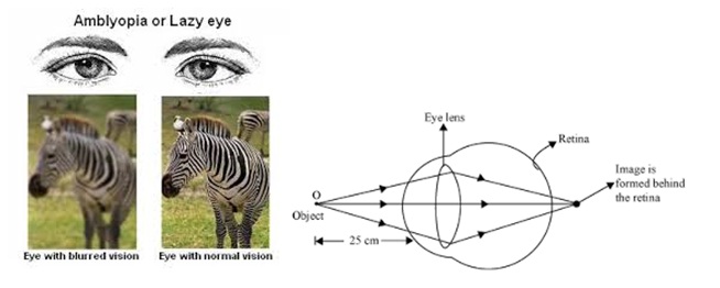
Amblyopia, also called lazy eye, is a disorder of sight due to the eye and brain not working well together. It results in decreased vision in an eye that otherwise typically appears normal. It is the most common cause of decreased vision in a single eye among children and younger adults.
The cause of amblyopia can be any condition that interferes with focusing during early childhood. This can occur from poor alignment of the eyes, an eye being irregularly shaped such that focusing is difficult, one eye being more nearsighted or farsighted than the other, or clouding of the lens of an eye. After the underlying cause is fixed, vision is not fully restored as the mechanism also involves the brain. Amblyopia can be difficult to detect and therefore vision testing is recommended for all children around the ages of four to five.
Many people with amblyopia, especially those who only have a mild form, are not aware they have the condition until tested at older ages, since the vision in their stronger eye is normal. People typically have poor stereo vision, however, since it requires both eyes. Those with amblyopia further may have, on the affected eye, poor pattern recognition, poor visual acuity, and low sensitivity to contrast and motion
Amblyopia is characterized by several functional abnormalities in spatial vision, including reductions in visual acuity (VA), contrast sensitivity function (CSF), and vernier acuity as well as spatial distortion, abnormal spatial interactions, and impaired contour detection. In addition, individuals with amblyopia suffer from binocular abnormalities such as impaired stereoacuity (stereoscopic acuity) and abnormal binocular summation. Also, a crowding phenomenon is present. These deficits are usually specific to the amblyopic eye. However, sub-clinical deficits of the "better" eye have also been demonstrated.Treatment of strabismic or anisometropic amblyopia consists of correcting the optical deficit (wearing the necessary spectacle prescription) and often forcing use of the amblyopic eye, by patching the good eye, or instilling topicalatropine in the good eye, or both.
Presbyopia is a natural occurrence where near vision becomes blurred, making it hard to focus while doing things like reading, using a mobile phone, or working on the computer. It is not a disease or illness; in fact, it is very common with age.
In young people, the eye’s lens is soft and flexible, readily changing shape to see images from different distances. As you age, the crystalline lens in your eye hardens and loses elasticity. With this loss of flexibility, your eyes are less able to adjust properly to focus near objects.
People commonly mistake the symptoms of presbyopia for longsightedness. However, the two conditions have different causes: longsightedness is a result of a misshapen cornea, whereas presbyopia is due to the loss of flexibility in the lens.
The telltale symptom of presbyopia is blurred vision while reading, sewing, using a mobile phone, or doing anything that requires near vision.
There are many options for people with presbyopia, including contact lenses. Recent technologies allow people who are entering into presbyopia to continue wearing contact lenses, instead of having to switch to bifocals, or reading glasses.
Common treatments for presbyopia include:
• Magnifiers
• Bifocal or varifocal spectacles
• Reading glasses
Near-Sightedness, also known as short-sightedness and myopia, is a condition of the eye where light focuses in front of, instead of on, the retina. This causes distant objects to be blurry while close objects appear normal. Other symptoms may include headaches and eye strain. Severe near-sightedness increases the risk of retinal detachment, cataracts, and glaucoma.
The underlying cause is believed to be a combination of genetic and environmental factors. Risk factors include doing work that involves focusing on close objects, greater time spent indoors, and a family history of the condition. It is also associated with a high socioeconomic class. The underlying mechanism involves the length of the eyeball being too long or less commonly the lens being too strong. It is a type of refractive error. Diagnosis is by eye examination.
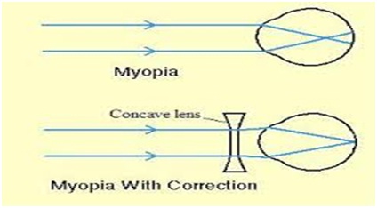
There is tentative evidence that near-sightedness can be prevented by having young children spend more time outside. This may be related to natural light exposure. Near-sightedness can be corrected with eyeglasses, contact lenses, or surgery. Eyeglasses are the easiest and safest method of correction. Contact lenses can provide a wider field of vision; however are associated with a risk of infection. Refractive surgery permanently changes the shape of the cornea.Myopia presents with blurry distance vision, but generally gives good near vision. In high myopia, even near vision is affected as objects must be extremely close to the eyes to see clearly, and people with myopia cannot read without their glasses prescribed for distance.
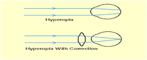
Far-Sightedness, also known as long-sightedness and hyperopia, is a condition of the eye in which light is focused behind, instead of on, the retina. This causes close objects to be blurry, while far objects may appear normal. As the condition worsens, objects at all distances may be blurred.] Other symptoms may include headaches and eye strain. People with hyperopia can also experience accommodative dysfunction, binocular dysfunction, amblyopia, and strabismus.
The cause is an imperfection in the eye: often when the eyeball is too short, or the lens cannot become round enough. It is a type of refractive error .
Correction is usually achieved by the use of convex corrective lenses. For near objects, the eye has to accommodate even more. Depending on the degree of hyperopia and the age of the person, which directly relates to the eye's accommodative ability, the symptoms can be different.
Far-sightedness primarily affects young children, with rates of 8% at 6 years and 1% at 15 years.The signs and symptoms of far-sightedness are blurry vision, headaches, and eye strain. The common symptom is eye strain. Difficulty seeing with both eyes (binocular vision) may occur, as well as difficulty with depth perception.As hyperopia is the result of the visual image being focused behind the retina, it has two main causes:
Low converging power of eye lens because of weak action of ciliary muscles.Abnormal shape of the cornea.
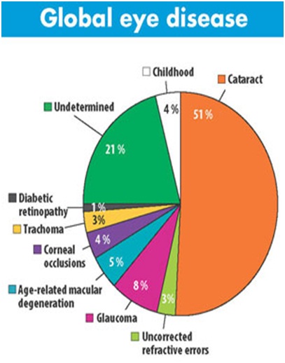
Cataract is a clouding of the lens in the eye which leads to a decrease in vision. Cataracts often develop slowly and can affect one or both eyes. Symptoms may include faded colors, blurry vision, halos around light, trouble with bright lights, and trouble seeing at night.This may result in trouble driving, reading, or recognizing faces. Poor vision caused by cataracts may also result in an increased risk of falling and depression.Cataracts are the cause of half of blindness and 33% of visual impairment worldwide.
Cataracts are most commonly due to aging but may also occur due to trauma or radiation exposure, be present from birth, or occur following eye surgery for other problems. Risk factors include diabetes, smoking tobacco, prolonged exposure to sunlight, and alcohol. Either clumps of protein or yellow-brown pigment may be deposited in the lens reducing the transmission of light to the retina at the back of the eye. Diagnosis is by an eye examination.

Prevention includes wearing sunglasses and not smoking. Early on the symptoms may be improved with glasses. If this does not help, surgery to remove the cloudy lens and replace it with an artificial lens is the only effective treatment. Surgery is only needed if the cataracts are causing problems and generally results in an improved quality of life. Cataract surgery is not readily available in many countries, which is especially true for women, those living in rural areas, and those who do not know how to read.
About 20 million people are blind due to cataracts. It is the cause of approximately 5% of blindness in the United States and nearly 60% of blindness in parts of Africa and South America. Blindness from cataracts occurs in about 10 to 40 per 100,000 children in the developing world, and 1 to 4 per 100,000 children in the developed world. Cataracts become more common with age. More than half the people in the United States had cataracts by the age of 80.
Glaucoma is a group of eye diseases which result in damage to the optic nerve and vision loss. The most common type is open-angle glaucoma with less common types including closed-angle glaucoma and normal-tension glaucoma. Open-angle glaucoma develops slowly over time and there is no pain. Side vision may begin to decrease followed by central vision resulting in blindness if not treated. Closed-angle glaucoma can present gradually or suddenly. The sudden presentation may involve severe eye pain, blurred vision, mid-dilated pupil, redness of the eye, and nausea. Vision loss from glaucoma, once it has occurred, is permanent.
Risk factors for glaucoma include increased pressure in the eye, a family history of the condition, migraines, high blood pressure, and obesity. For eye pressures a value of greater than 21 mmHg or 2.8 kPa is often used with higher pressures leading to a greater risk. However, some may have high eye pressure for years and never develop damage. Conversely, optic nerve damage may occur with normal pressure, known as normal-tension glaucoma. The mechanism of open-angle glaucoma is believed to be slow exit of aqueous humor through the trabecular meshwork while in closed-angle glaucoma the iris blocks the trabecular meshwork. Diagnosis is by a dilated eye examination. Often the optic nerve shows an abnormal amount of cupping. If treated early it is possible to slow or stop the progression of disease with medication, laser treatment, or surgery. The goal of these treatments is to decrease eye pressure. A number of different classes of glaucoma medication are available. Laser treatments may be effective in both open-angle and closed-angle glaucoma. A number of types of glaucoma surgeries may be used in people who do not respond sufficiently to other measures. Treatment of closed-angle glaucoma is a medical emergency.
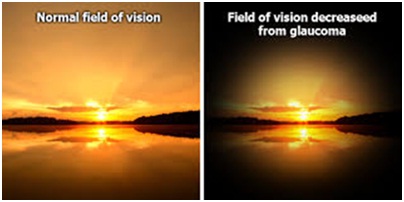
About 11 to 67 million people have glaucoma globally. The disease affects about 2 million people in the United States. It occurs more commonly among older people. Closed-angle glaucoma is more common in women. Glaucoma has been called the "silent thief of sight" because the loss of vision usually occurs slowly over a long period of time.Worldwide, glaucoma is the second-leading cause of blindness after cataracts. The word "glaucoma" is from ancient Greek glaukos which means blue, green, or gray. In English, the word was used as early as 1587 but did not become commonly used until after 1850, when the development of the ophthalmoscope allowed people to see the optic nerve damage.
Diabetic Retinopathy, also known as diabetic eye disease, is when damage occurs to the retina due to diabetes. It can eventually lead to blindness.It is an ocular manifestation of diabetes, a systemic disease, which affects up to 80 percent of all patients who have had diabetes for 20 years or more. Despite these intimidating statistics, research indicates that at least 90% of these new cases could be reduced if there were proper and vigilant treatment and monitoring of the eyes. The longer a person has diabetes, the higher his or her chances of developing diabetic retinopathy.
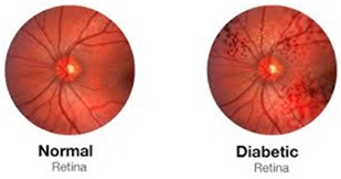
Each year in the United States, diabetic retinopathy accounts for 12% of all new cases of blindness. It is also the leading cause of blindness for people aged 20 to 64 years.Diabetic retinopathy often has no early warning signs. Even macular edema, which can cause rapid vision loss, may not have any warning signs for some time. In general, however, a person with macular edema is likely to have blurred vision, making it hard to do things like read or drive. In some cases, the vision will get better or worse during the day.
Macular Degeneration, also known as age-related macular degeneration (AMD or ARMD), is a medical condition which may result in blurred or no vision in the center of the visual field. Early on there are often no symptoms. Over time, however, some people experience a gradual worsening of vision that may affect one or both eyes. While it does not result in complete blindness, loss of central vision can make it hard to recognize faces, drive, read, or perform other activities of daily life. Visual hallucinations may also occur but these do not represent a mental illness.
Macular degeneration typically occurs in older people. Genetic factors and smoking also play a role. It is due to damage to the macula of the retina. Diagnosis is by a complete eye exam. The severity is divided into early, intermediate, and late types.[1] The late type is additionally divided into "dry" and "wet" forms with the dry form making up 90% of cases.
Prevention includes exercising, eating well, and not smoking. Antioxidant vitamins and minerals do not appear to be useful for prevention. There is no cure or treatment that returns vision already lost. In the wet form, anti-VEGF medication injected into the eye or less commonly laser coagulation or photodynamic therapy may slow worsening. Supplements in those who already have the disease may slow progression.
In 2010 it affected 23.5 million people globally. In 2013 moderate to severe disease affected 13.4 million and it is the fourth most common cause of blindness after cataracts, preterm birth, and glaucoma.. It most commonly occurs in people over the age of fifty and in the United States is the most common cause of vision loss in this age group. About 0.4% of people between 50 and 60 have the disease, while it occurs in 0.7% of people 60 to 70, 2.3% of those 70 to 80, and nearly 12% of people over 80 years old
Scribd
Slideshare
Webmd
Youtube
World Health Organisation
Kids Health
The Human Body Book By Webster